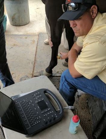
Many structures in the body, such as ligaments and tendons, do not show up well on radiographs, but can be examined very well with ultrasound. In an ultrasound examination, high frequency sound waves are generated and aimed at a structure. Some of these waves are bounced back by the structure being examined and a digital image is produced from these echoed waves. Other structures, such as the heart and lungs, can’t be radiographed in the adult horse because of size, but can be visualized very well with ultrasound. Ultrasound examinations gather valuable information in cases of pneumonia and colic, among others.
The ultrasound machine we use at Temecula Creek Equine is of a laptop design, so it is very portable and can be used in your barn for a true “horse side” examination. The examination can be recorded and stored for playback at another time. This allows review of the information as well as transmitting to another veterinarian for a consultation, when desired.
Ultrasound is also used extensively in reproduction. It gives us much information about uterine health and follicle development to aid in breeding. It is the primary method of pregnancy testing and can detect an embryo as early as 15 days of age, detect twins and monitor fetal health.